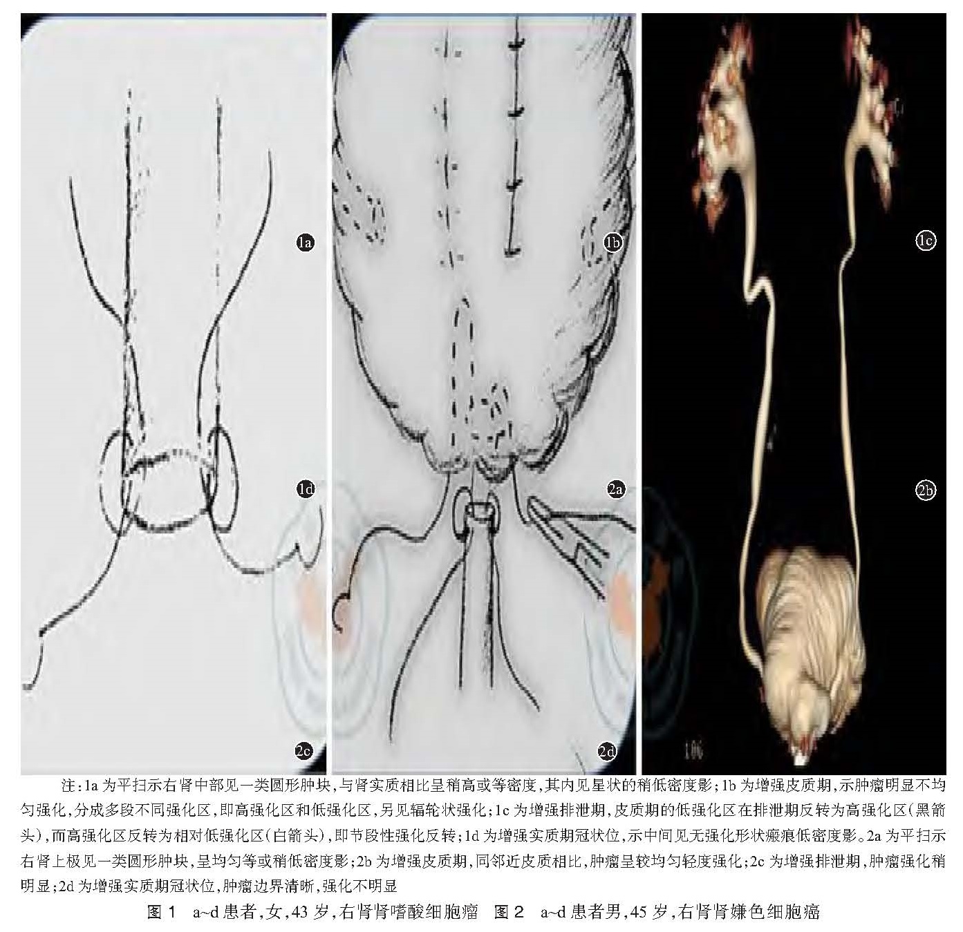
目的 初步探讨肾嗜酸细胞腺瘤( RO)和嫌色细胞癌( CCRC)的多层螺旋 CT影像学特征的鉴别诊断价值。方法 回顾性分析 2003年 6月至 2015年 12月我院经手术病理证实的 9例肾嗜酸细胞腺瘤和 12例嫌色细胞癌的患者资料,术前全部行螺旋 CT平扫及三期动态增强,并分析其影像学特征。结果 3例( 33.3%)RO病灶可见钙化,而 1例( 8.3%)CCRC病灶可见钙化,它们之间差异无统计学意义( P>0.05)。5例( 55.6%)RO病灶、1例( 8.3%)CCRC病灶见星状或条索状纤维瘢痕;6例(66.7%)RO病灶、2例( 16.7%)CCRC病灶出现辐轮状强化; 6例( 66.7%)RO病灶、2例( 16.7%)CCRC病灶出现节段性强化反转,它们之间差异均有统计学意义( P<0.05);所有患者均未出现腹膜后淋巴结肿大。3例( 33.3%)RO病灶、9例( 75.0%)CCRC病灶显示均匀性强化;7例 (77.8%)RO病灶、2例(16.7%)CCRC病灶显示明显强化,它们之间差异均有统计学意义( P<0.05)。CT平扫,RO病灶 CT值高于 CCRC病灶,CT动态增强各期相,RO病灶 CT值也高于 CCRC病灶。结论 星状或条索状的纤维瘢痕、辐轮状强化、节段性强化反转、明显性强化更多出现在 RO患者中,而均匀性强化更多出现在 CCRC患者中,术前多层螺旋 CT有助于鉴别 RO和 CCRC。
Objective To compare the multi-slice computed tomography (MSCT) characteristics of renal oncocytoma (RO) and chomophobe cell renal carcinoma (CRCC). Methods From June 2003 to December 2015, dinical data of 9 patients with RO and 12 patients with CRCC were studied retrospectively in the Third Affiliated Hospital of Sun Yat-sen University. MSCT was performed to investigate tumor imaging characteristics in plain and 3-phase dynamic enhanced scans. Results Calcifications were observed in 3 (33.3%) patients with RO and in 1 (8.3%) patients with CCRC, there was no significant difference between RO and CCRC(P>0.05). five (55.6%) patients with RO had stellate or funicular scars as did one (8.3%) patient with CCRC. Spoken-wheel-like enhancement was visible in 6 (66.6%) patients with RO and in 2(16.7%) with CCRC. six (66.7%) patients with RO and 2 (16.7%) patients with CCRC showed segmental inversion. Combined evaluation of stellate or funicular scar, spoken-wheel-like enhancement, segmental enhancement inversion and obvious enhancement features had significant differences between RO and CCRC (P<0.05). No patients with RO and CCRC had retroperitoneal lymph node enlargement. Three (33.3%) patients with RO and 9 (75.0%) patients with CCRC showed homogeneous enhancement. Seven (77.8%) patients with RO and 2 (16.7%) patients with CCRC showed obvious enhancement. There was significant difference between RO and CCRC (P<0.05). The attenuation of RO tumors was greater than that of CCRC tumors on unenhanced CT. Enhancement was higher with RO than with CCRC tumors in all phases. Conclusion CT imaging features such as stellate or funicular scar, spoken-wheel-like enhancement, segmental enhancement inversion, and obvious enhancement were more common in RO, but homogeneous enhancement was more common in CCRC and they may help in differentiating RO from CCRC.
![表1 肾脏肾嗜酸细胞瘤(RO)和肾嫌色细胞癌(CCRC)的CT 主要影像学特征比较[(例,%)]<br/>](2016年06期/pic15.jpg)